The Ultrastructure of Ocelli of a Pea Aphid Acanthosiphon pisum (Harris).
The Ultrastructure of Ocelli of a Pea Aphid Acanthosiphon pisum (Harris).
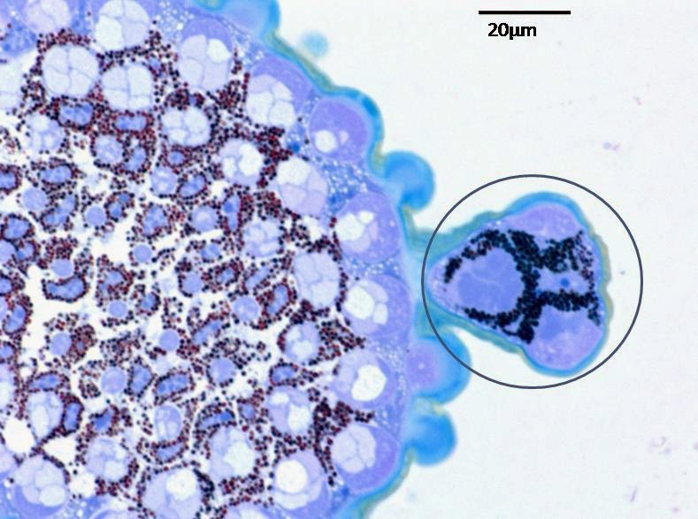


The morphology and fine structure of the ocelli of Acanthosiphon pisum have been analyzed by means of light and electron microscopy. The three ocelli of this species are located near the compound eyes. Externally, the ocelli are marked by the corneal lenses virtually spherical in form. A single layer of corneagen cells lies below the cuticular lens. The corneagen and retinal cells are arranged in a cup-like manner below the cuticular lens. Each retinal cell contribute their microvilli to form rhabdom. Below the rhabdom the oviform nuclei of the retinal cells appear. Screening pigment granules are present within the retinal cell. Spherical mitochondria are homogeneously distributed in the cytoplasm of the cell body. At the proximal region retinal cells form axon and exit the basal lamina.


Comments
Post a Comment